Advancements in Imaging Techniques for Eye Care
Have you ever wondered how doctors see inside your eyes without actually touching them? Thanks to exciting advancements in imaging techniques, eye care has reached new heights. These technologies help detect problems early and provide better treatment. Lets dive into the world of eye imaging and see how it makes a difference for our vision.
What Are Imaging Techniques in Eye Care?

Imaging techniques are tools that help eye care professionals look at the inside of your eyes. They can spot diseases like glaucoma or macular degeneration much earlier than before. These technologies include various methods such as optical coherence tomography (OCT) and fundus photography.
Think of imaging techniques like taking a picture. Just as a camera captures an image, these methods capture detailed pictures of your eyes. This allows doctors to view the eye’s structures, just as a mechanic might check a car engine.
Why Are These Advancements Important?

The importance of advanced imaging techniques cannot be overstated. According to the World Health Organization, around 2.7 billion people suffer from vision problems worldwide. Early detection can lead to better outcomes and even save vision.
- Early Diagnosis: Detect issues before they become serious.
- Better Treatment: Tailor treatments based on precise images.
- Monitoring Progress: Track changes in eye health over time.
What Are the Latest Imaging Techniques?

Lets look at some of the latest and coolest imaging techniques that eye doctors use today.
1. Optical Coherence Tomography (OCT)
OCT is like a super-smart camera for your eyes. It uses light waves to take cross-section images of your retina. This high-resolution imaging helps doctors see layers of your retina. it’s essential for diagnosing conditions like diabetic retinopathy and macular holes.
Think of it like slicing a loaf of bread. Each slice shows a different layer, helping you understand the whole loaf. With OCT, doctors can pinpoint exactly where a problem lies.
2. Fundus Photography
Fundus photography captures detailed images of the back of your eye, where the retina resides. This technique provides a broad view of the retinal surface and blood vessels. it’s crucial for monitoring diseases like glaucoma and age-related macular degeneration.
Imagine taking a wide-angle photo of a landscape. You can see everything at once. Fundus photography does the same for your eye.
3. Slit-Lamp Imaging
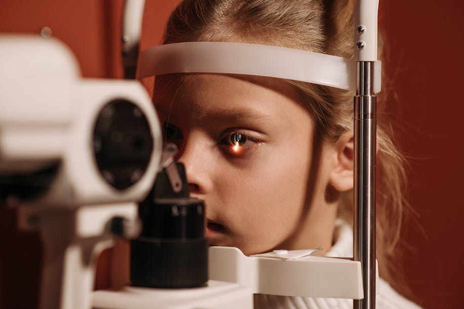
The slit lamp is a common tool that combines a microscope and a light source. It allows eye doctors to examine the front structures of the eye in detail. With new imaging technologies, slit-lamp systems can now capture high-quality images, providing more information about conditions like cataracts and corneal diseases.
This is like using a magnifying glass to examine a small object up close. You get a crystal-clear view of details that are otherwise missed.
4. Ultrasonography
Ultrasonography uses sound waves to create images of the eye. it’s particularly useful for examining the eye’s interior and detecting tumors or other abnormalities. This method is safe, quick, and painless.
Picture this as a sonar system. Just as a dolphin uses sound to navigate underwater, this technique helps doctors see what lies beneath the surface of the eye.
How Do These Techniques Benefit Patients?

Modern imaging techniques offer numerous benefits for patients. Here are a few key advantages:
- Comfort: Most imaging procedures are quick and non-invasive.
- Speed: Results can often be available the same day.
- Precision: Higher accuracy in diagnosing eye conditions.
Are There Risks Involved?
Generally, these imaging techniques are very safe. However, some patients may feel discomfort during certain procedures, like dilating eye drops used in fundus photography. It’s essential to discuss any concerns with your eye doctor.
Always remember, the benefits of early detection and accurate diagnosis far outweigh potential risks.
what’s Next in Eye Imaging Technology?
Innovation never stops! Researchers are continually working on improving imaging techniques. One exciting area is artificial intelligence (AI). AI can analyze images faster and with greater accuracy than the human eye. This could lead to quicker diagnoses and better treatment plans.
Imagine having a computer assistant that helps your doctor make decisions or even suggests treatments based on what it sees. This is the future of eye care!
Common Questions About Eye Imaging Techniques
Many people have questions about these technologies. Lets address some of the most common ones.
Are imaging techniques painful?
No, most imaging techniques are painless and non-invasive. Some may require eye drops for dilation, which can cause temporary discomfort.
How often should I have my eyes imaged?
Your eye doctor can recommend how often you should undergo imaging based on your health and risk factors. Regular check-ups are essential, especially if you have a history of eye problems.
Can imaging techniques completely replace eye exams?
No, imaging techniques complement eye exams but do not replace them. Regular eye exams are crucial for overall eye health.
Take Action for Your Eye Health
Understanding these advancements in imaging techniques is vital for maintaining good eye health. Schedule regular appointments with your eye doctor to take advantage of these technologies. don’t wait until you notice a problem!
For more detailed information on eye care, check out the American Academy of Ophthalmology.
In conclusion, advancements in imaging techniques are transforming eye care. They allow for early detection, better treatment options, and improved patient outcomes. Stay informed and proactive about your eye health!


